Researchers link breast cancer and bone growth
Interdisciplinary breast cancer research
A research team consisting of materials scientists from the Max Planck Institute of Colloids and Interfaces (MPICI) in Potsdam and biologists from Cornell University in Ithaca, USA revealed that bones may grow in response to certain signals from a distant breast tumor. This may be a preemptive defense mechanism against skeletal metastasis. These study results could influence diagnostic tests and therapeutic breast cancer treatments in the future.
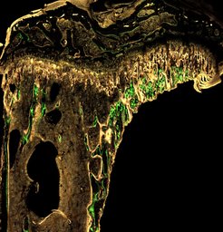
; method: dynamic histomorphometry")
Breast cancer is the most common cancer in women, and bones are a prime target for breast cancer metastasis. The mechanisms that influence the development of skeletal metastasis remain poorly understood. What is known is that tumor cells emit various signaling molecules, making organs vulnerable to the spread of cancer. It is also known that breast cancer with skeletal metastasis causes bone degradation, as metastatic tumors activate bone-degrading cells and inhibit bone-forming cells.
Led by Prof. Dr. Peter Fratzl, the Potsdam materials scientists made a surprisingly opposite discovery: "Using an animal model developed by Cornell University, we found that early-stage bones grow faster when exposed to tumor signaling molecules. This happens by adding more layers of mineralized tissue in a short period of time," says Peter Fratzl, Director of the Department of Biomaterials at MPICI. This observation could potentially be a defense mechanism of the body trying to prevent metastasis. Such a protective mechanism could find application in diagnostics as well as in the development of therapeutic treatments. "Overall, this interdisciplinary study demonstrates how engineering and physical sciences approaches are needed to really understand the full spectrum of changes that tumors might introduce to an organism," added Prof. Claudia Fischbach, co-director of the Cornell Center on the Physics of Cancer Metabolism, New York.
The method
For their studies of the structure and composition of the bone microenvironment, the Potsdam research team used a number of high-resolution imaging techniques: Micro-computed tomography to study the bone architecture, Raman imaging to scan bones’ chemical spectra, small-angle X-ray scattering (SAXS) to reveal mineral bone properties. The main technique used was dynamic histomorphometry, in which fluorescent dyes are integrated into the bone at different intervals. This creates time stamps showing the rate of bone formation.
The interdisciplinary and international research project was supported by the Human Frontier Science Program with a three-year grant of 1 million US Dollars.


