
Soft Matter Electron Microscopy
Overview of EM facilities at the Sustainable and Bio-inspired Materials Department
Our EM facilities are currently under development and will soon house a cryo transmission electron microscope (cryoTEM) and a cryo ion-beam scanning electron microscope (cryoFIB/SEM), complementing the existing facilities at MPIKG. These facilities are dedicated to the structural characterization of radiation-sensitive soft and biological materials.
 Chitin microfibrils of Phaeocystis globosa (B) Epidermal cells of Viburnum tinus fruit (C) Electron diffraction diagram of synthetic oligosaccharide crystal (E) Lattice image of chitin microfibril cross section (F) Cellulose nanocrystals")
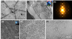
(A) Chitin microfibrils of Phaeocystis globosa (B) Epidermal cells of Viburnum tinus fruit (C) Electron diffraction diagram of synthetic oligosaccharide crystal (E) Lattice image of chitin microfibril cross section (F) Cellulose nanocrystals
Available Equipment
Transmission Electron Microscopy
JEOL JEM F-200 “Cryo” (available in March 2025)
- Cold field emission gun
- Cryo polepiece for low ice contamination rate
- Accelerating voltage: 60, 80, 120, and 200 kV.
- TEM lattice resolution at 200 kV: 0.1 nm
- STEM resolution (HAADF): 0.14 nm
- α-tilt angle: ±80°
- Hole-free phase plate
- Gatan Metro In-situ direct electron detector
- JEOL STEM detector
- Gatan Elsa cryo-transfer holder (698.ULP) with α-tilt angle > ±70°
Cryo Focused ion beam / scanning electron microscopy
- Volume imaging
- TEM lamella preparation at room and cryo temperature (lift-out, on-grid)
- Chemical analytical capability
Scanning Electron Microscopy
Tescan Clara Field-emission SEM
- Schottky emitter
- Accelerating voltage: 50 V-30 kV
- In-chamber Everhart-Thornley detector
- In-chamber BSE detector
- In-beam multi-detector
- EDS detector (coming soon)
- Beam deceleration function