Home News Research Publications Group Join us


DIMOVA LAB

Biomembranes and more

Giant lipid vesicles are a fascinating model membrane system with increasing popularity. Their dimensions are in the cell-size range. Compared to conventional vesicles with sizes in the range of hundred nanometers, these giants are a convenient tool for direct microscopy observation of phenomena occurring at the cell-size scales. One can observe them under phase contrast, differential interference contrast (DIC), conventional fluorescence and confocal microscopy. The three reviews from our group (see links below) and an The Giant Vesicle Book (Eds. R. Dimova & C. Marques, CRC Press, 2019) describe some research based on the use of giant vesicles:

A practical guide to giant vesicles. Probing the membrane nanoregime via optical microscopy, R. Dimova et al. J. Phys.: Condens. Matter 18, S1151-S1176 (2006) [Full text]

Giant vesicles: a biomimetic tool for membrane characterization, R. Dimova, in Advances in Planar Lipid Bilayers and Liposomes, vol. 16, pp. 1-50 (2012) [For full text access, send me an email]

Giant vesicles and their use in assays for assessing membrane phase state, curvature, mechanics and electrical properties, R. Dimova, Annu. Rev. Biophys. 48, 93-119 (2019) [Full text]

A few words about giant vesicles

a vesicle enclosing aqueous droplets (DIC)


Read more

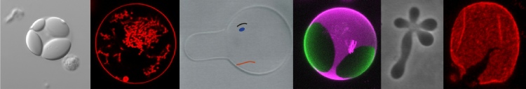
a vesicle with internal tubes (confocal cross section)

Read more

a tough diblock-copolymer vesicle after being released from a micropipette (DIC)

Read more

a multicomponent vesicle with domains (3D confocal projection)

Read more

a palm-like vesicle

a wrinkled gel-phase vesicle after being crack-opened

Read more

Last modified: 13-Nov-20 12:36:29 PM

Imprint