Development of Mineralized Skeletal Materials
The aim of our research group is to understand the structural and functional properties of the materials surrounding the cells and the mineralized matrix during the development of skeletal tissues.
The materials in which cells reside is called the extracellular matrix (ECM), a complex entity composed of macromolecules that act as a regulator and an information repository for the cellular functions. In addition, the ECM provides structural and mechanical support to the cells and the tissue, playing a crucial role during growth, development, morphogenesis as well as the maintenance of the tissue. Yet, the role and the physical mechanisms by which the ECM regulates cell behavior and skeletal tissue organization remains elusive. To decipher these mechanisms, we use a structural approach to gain insight into the 3D spatial arrangement and structure of the different components of skeletal material.
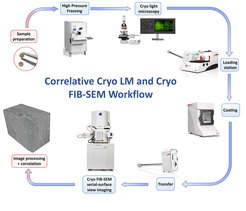
We use imaging techniques covering multiple length-scales from the micrometer to the nanometer level (light microscopy of histological sections, X-ray microtomography, electron microscopy) combining both 2D and 3D imaging to obtain a comprehensive analysis of skeletal tissues. To gain access to the ultrastructural level in 3D, we are developing a methodology combining the Focused Ion Beam- Scanning Electron Microscope (FIBSEM) with the correlative light and electron microscopy (CLEM) system in cryo-condition. This approach allows to image a large volume at high resolution in correlation with the localization of fluorescent macromolecules in order to better understand the principles governing skeletal material formation by visualizing cells and tissues in their close to native state.